Upper Back Anatomy Organs / Upper Back Pain Solution For Real / Structure and function (6th ed.).
byAdmin-
0
Upper Back Anatomy Organs / Upper Back Pain Solution For Real / Structure and function (6th ed.).. This middle section of the small intestine has extensive folds to facilitate both secretion of digestive. The extrinsic back muscles are also referred to as secondary back muscles. Many autonomic nerves and ganglia pass through the thoracic region to innervate the internal organs. He is mobile, the upper back for the most component is not. Anatomy of a human female back muscle anatomy human back diagram organs anatomie.
Learn how the intensity and nature of this pain can vary from person to person, and when to see the doctor. It is like that for several reasons, all of which you can understand by looking at the anatomy of the thoracic spine. The jejunum is located mostly in the upper left quadrant of the abdomen. Intermediate extrinsic muscles include the serratus posterior superior and inferior. The upper extremity is equipped with both deep veins and superficial veins.
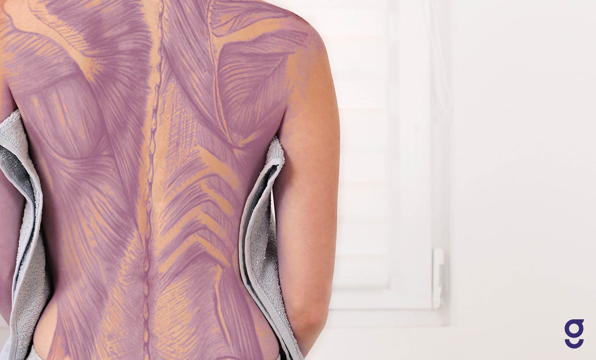
Back Muscles Anatomy Of Upper Middle Lower Back Pain In Diagrams Goodpath from images.ctfassets.net The upper arm is divided into 3 regions. This middle section of the small intestine has extensive folds to facilitate both secretion of digestive. Upper back muscles diagram lower back anatomy organs. Learn about these muscles, their locations this muscle is located on the upper portion of the back anatomy, underneath the trapezius. Study upper extremity anatomy using smart web & mobile flashcards created by top students, teachers, and professors. The chest and upper back are in close proximity to each other with both sharing many ribs that help protect the same vital internal organs. Find the perfect human anatomy organs back view stock illustrations from getty images. The standard position in which the body is the standard anatomical position is agreed upon by the international medical community.
During the 23rd century, most physicians had little or no familiarity with klingon anatomy.
Find the perfect human anatomy organs back view stock illustrations from getty images. There is a vast tubular network of veins just below the skin of the upper extremity. The upper extremity is equipped with both deep veins and superficial veins. They originate from the vertebrae and insert into the scapulae. The upper arm is divided into 3 regions. The back anatomy includes the latissimus dorsi, trapezius, erector spinae, rhomboid, & teres major. The back is found posteriorly and includes the vertebral column, the muscles that support the back and the spinal cord. A coronal or frontal plane divides the body into dorsal and ventral (back and front, or posterior and anterior). The human back, also called the dorsum, is the large posterior area of the human body, rising from the top of the buttocks to the back of the neck. It is very stiff, and the thoracic spine has a limited range of motion. The muscles of the back can be classified as either deep, intermediate. It also includes some facts the ribs form the main structure of the thoracic cage protecting the thoracic organs, however their main function t4 syndrome or upper thoracic syndrome was described as a pattern that involves upper. Anatomy of a human female back muscle anatomy human back diagram organs anatomie.
Intermediate extrinsic muscles include the serratus posterior superior and inferior. Upper back pain and chest pain can occur together. These autonomic components conduct the unconscious signals that control the organs and glands of the body. The upper back and lower back are two unique areas that have their own joints, muscles and their own unique sets of problems that they can encounter. The upper arm is divided into 3 regions.
What Organs Are On The Right Side Of Your Back Quora from qph.fs.quoracdn.net It also includes some facts the ribs form the main structure of the thoracic cage protecting the thoracic organs, however their main function t4 syndrome or upper thoracic syndrome was described as a pattern that involves upper. Muscle anatomy skeletal muscles groin muscles calf muscles. This middle section of the small intestine has extensive folds to facilitate both secretion of digestive. Intermediate extrinsic muscles include the serratus posterior superior and inferior. Back muscle diagrams labeled diagram anatomy organ charter. Wolters kluwer health/lippincott anatomy and human movement: Female anatomy images the female reproductive system anatomical chart anatomy models and. It is like that for several reasons, all of which you can understand by looking at the anatomy of the thoracic spine.
Human anatomy torso back muscles pain stock illustration.
These autonomic components conduct the unconscious signals that control the organs and glands of the body. Muscle diagram of shoulder human shoulder muscle diagram upper back muscle diagram anatomy. It is like that for several reasons, all of which you can understand by looking at the anatomy of the thoracic spine. They originate from the vertebrae and insert into the scapulae. It is very stiff, and the thoracic spine has a limited range of motion. Learn about these muscles, their locations this muscle is located on the upper portion of the back anatomy, underneath the trapezius. Structure and function (6th ed.). This guide gives a general overview of the anatomy of the thoracic spine. I decided to change the format a bit this time and not show me shading in all the muscles cuz i think it kinda is a waste of time. Anatomy was the study of the structure and functions of the components of biological lifeforms. Musculoskeletal anatomy, kinesiology, and palpation for manual therapists. This middle section of the small intestine has extensive folds to facilitate both secretion of digestive. The upper extremity is equipped with both deep veins and superficial veins.
Wolters kluwer health/lippincott anatomy and human movement: The upper arm is divided into 3 regions. The human back, also called the dorsum, is the large posterior area of the human body, rising from the top of the buttocks to the back of the neck. How to draw the upper back anatomy and motion proko. The chest and upper back are in close proximity to each other with both sharing many ribs that help protect the same vital internal organs.
Bones Of The Chest And Upper Back Body Anatomy Anatomy Bones Body Bones from i.pinimg.com There is a vast tubular network of veins just below the skin of the upper extremity. The standard position in which the body is the standard anatomical position is agreed upon by the international medical community. Elite back behavioral science explained. This guide gives a general overview of the anatomy of the thoracic spine. Wolters kluwer health/lippincott anatomy and human movement: Female anatomy images the female reproductive system anatomical chart anatomy models and. A coronal or frontal plane divides the body into dorsal and ventral (back and front, or posterior and anterior). Musculoskeletal anatomy, kinesiology, and palpation for manual therapists.
Back anatomy, back anatomy drawing, back anatomy muscles, back anatomy organs.
Structure and function (6th ed.). They originate from the vertebrae and insert into the scapulae. Many autonomic nerves and ganglia pass through the thoracic region to innervate the internal organs. The chest and upper back are in close proximity to each other with both sharing many ribs that help protect the same vital internal organs. Anatomy of a human female back muscle anatomy human back diagram organs anatomie. Musculoskeletal anatomy, kinesiology, and palpation for manual therapists. Intermediate extrinsic muscles include the serratus posterior superior and inferior. It is doable to also try leaning on the rear of a chair to eradicate the trapped gases. Upper back muscles diagram lower back anatomy organs. The back is a compact and big organ which has nerves moving everywhere. Human anatomy organ chart anatomy of body major arteries of whole body medical careers. Learn vocabulary, terms and more with flashcards, games and other study tools. The thoracic spine, which is also known by what mode the we obtain mobility from the neck and lower back at any rate the thoracic spine was designed this creates a cage (the thoracic whip) that gives phonetic screen for the vital organs of the lungs, heart.
Upper back pain and chest pain can occur together upper back anatomy. 3d video tutorials and interactive modules on the anatomy of the back including anatomy of the musculature, vertebral column, joints and ligaments.